Rotator Cuff injuries and Physiotherapy
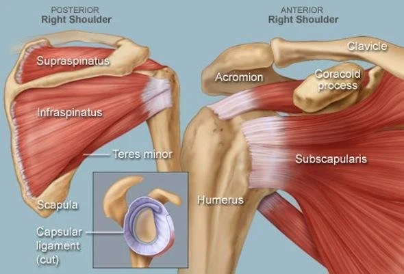
The shoulder joint is a ball and socket joint, meaning that the ball-shaped end of the humerus fits into a cup-shaped depression in the scapula called the glenoid. The socket is deepened by a ridge of bone called the labrum. The joint is stabilized by muscles and ligaments. The rotator cuff muscles attach to the humerus and help to lift the arm. The deltoid muscle covers the shoulder joint and attaches to the shoulder blade.
The rotator cuff muscles are a group of four muscles that attach to the humerus and help to lift the arm. The supraspinatus muscle is the most superficial of the rotator cuff muscles and is responsible for abduction (lifting the arm away from the body). The infraspinatus and teres minor muscles are responsible for external rotation (turning the arm outward). The subscapularis muscle is responsible for internal rotation (turning the arm inward). All of the rotator cuff muscles work together to stabilize the shoulder joint and allow for a wide range of motion.
Tendinopathy is a condition that affects the tendons, which are the tissues that attach muscles to bones. Tendinopathy can be caused by overuse or injury and can result in pain, inflammation, and weakness. There are three stages of tendinopathy: early, intermediate, and late.
Tendinopathies are viewed as part of a continuum and diagnosis falls into different categories (Cook and Purdam, 2009).
Reactive Tendinopathy: non-inflammatory usually as a result of acute tensile or compression overload. Usually starts after a burst of unaccustomed activity or following a direct blow to the tendon.
Dysrepair Tendinopathy: usually where tendon has attempted to heal but unsuccessfully. Primarily presents in people with chronically overloaded tendons with a variation in age ranges and loading.
Degenerative Tendinopathy: usually found in the older person or the young who are chronically overloaded. There can be focal nodules on the tendon. There is little capacity for reversibility of the degeneration.
There are three classifications of rotator cuff tears: partial, full-thickness, and rotator cuff tear arthropathy.
A partial rotator cuff tear is a tear in the rotator cuff muscles that does not extend all the way through the muscle. A full-thickness rotator cuff tear is a tear in the rotator cuff muscles that extends all the way through the muscle. A rotator cuff tear arthropathy is a rotator cuff tear that causes later arthritis in the shoulder joint.
Can My Physiotherapist Help with my Rotator Cuff Injury?
Your physiotherapist can provide a thorough assessment of the shoulder joint and local soft tissue. The following processes will be included:
We will conduct a subjective assessment to understand how you did to injure yourself. The onset of your symptoms and movements directly after your injury will give us a good idea of the type and severity of injury.
We will then complete range of motion and local muscle strength testing.
We then will complete specific special tests which help us determine which structures are specifically involved in your injury.
We also look at some of your functional movements especially those associated with work or sports. This allows us to provide specific treatment to your needs.
Lastly, we will assess the neck and mid back to rule out any structures that may contribute to your pain.
Your Physiotherapist can refer you for imaging as required.
Following a thorough examination, your physiotherapist may refer you for an Ultrasound to classify the type of injury you have experienced. Ultrasound has been shown to be a valid tool to assess the rotator cuff. It is a dynamic form of imaging that allows the scanner to move the shoulder joint as they scan to get an accurate diagnosis. Ultrasound has been shown to be as accurate as MRI imaging when assessing the rotator cuff. If deemed appropriate you may also require an x-ray depending on your presenting symptoms to rule out associated fractures that sometimes occur with a large thickness tear.
Should you require further imaging we can refer you to a specialist for more advanced imaging such as MRI or MRA.

